Introduction
Cartilage damage is a common and frustrating issue, especially for those who enjoy sports or are experiencing the natural effects of aging. Because cartilage does not heal well on its own, finding effective treatment options is crucial to relieve pain and restore healthy joint movement . ChondroFiller is a pioneering solution: a one-step, collagen-based implant designed specifically to repair small areas of cartilage damage. In this article, we’ll explain how ChondroFiller works, what results you can expect, and why receiving expert clinical care makes all the difference in your recovery.
Understanding ChondroFiller
So, what is ChondroFiller ? In simple terms, it’s a sterile, cell-free implant made from type I collagen—the primary protein found in natural cartilage. Think of ChondroFiller as a biological scaffold that fills in the damaged area. This collagen matrix acts as a supportive framework, encouraging your body’s own stem cells to move into the implant. Once there, these cells begin producing the building blocks of healthy cartilage, helping to rebuild and restore the affected tissue.
ChondroFiller has been designed to closely mimic the way real cartilage responds to daily movement and pressure. Thanks to its unique mechanical properties, it doesn’t just fill the space but actively supports regeneration, moving and adapting as you do.
Clinical experts also highlight that ChondroFiller can be applied using minimally invasive techniques, making it a modern and less disruptive option for those needing cartilage repair .
How ChondroFiller Repairs Cartilage
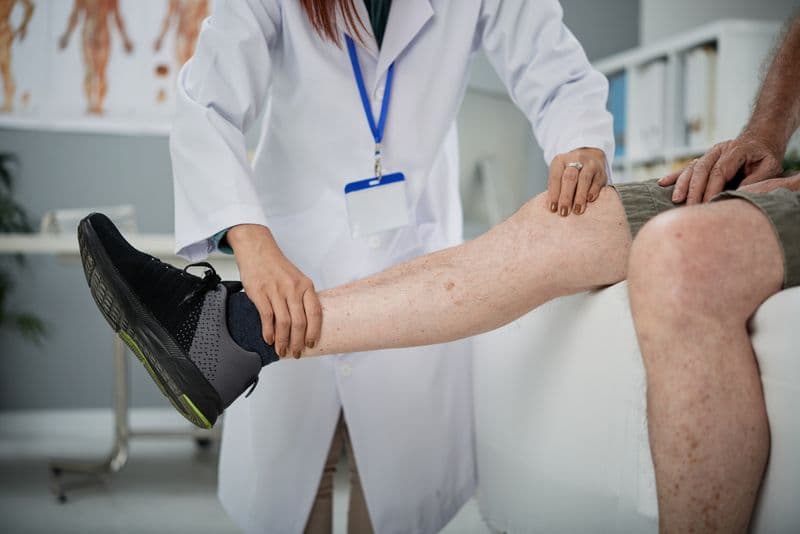
The ChondroFiller procedure is carried out during keyhole surgery (arthroscopy). Here’s what happens: after cleaning and drying the damaged cartilage area, the surgeon injects the warmed, gel-like ChondroFiller into the defect. The material then sets into a soft, flexible scaffold that perfectly fills the damaged spot. This scaffold encourages your own stem cells to migrate into the area, where they transform into cartilage-producing cells (chondrocytes). These cells begin repairing the tissue, gradually restoring healthy cartilage .
Studies and laboratory research have shown that ChondroFiller ’s adaptable, viscous structure offers excellent support for new tissue growth, potentially cushioning the joint as the repair takes place. The delivery technique, often using specialized surgical tools, also ensures the implant fills the defect precisely for optimal healing.
Procedure and Safety Profile
The typical ChondroFiller procedure starts with a diagnostic arthroscopy, allowing the surgeon to examine the joint and remove unstable cartilage edges. To help the implant adhere, the joint is gently dried before the warmed gel is injected. The gel sets within minutes. After surgery, the joint is immobilized briefly—usually in a plaster splint for about 48 hours—before beginning gentle movement. Patients use crutches and limit weight-bearing activities for about six weeks as part of a guided rehabilitation program.
ChondroFiller has a strong safety record. The procedure only requires one surgical step and avoids taking cartilage from another area of the body, reducing risks and recovery time. It’s performed in a professional clinical setting, customized for patient comfort and recovery.
Studies have found that ChondroFiller is safe and effective, with no complications reported in early clinical experiences. Research also emphasizes that matching the mechanical properties of natural cartilage is a key reason for ChondroFiller ’s success in joint repair.
Evidence from Clinical Studies
What does the research say? Clinical studies have shown very encouraging results. For example, in a study of 44 patients with cartilage defects in the knee or ankle, about 80% reported good or very good results and said they would have the procedure again. On average, patients saw significant improvements in function, and none reported their symptoms worsening.
Other research comparing ChondroFiller to standard treatments like microfracture found that ChondroFiller led to excellent filling of cartilage defects and integrated well with existing tissue up to 12 months after surgery. These findings point to ChondroFiller as a minimally invasive alternative that produces strong outcomes for patients.
Surgeons have also noted that precise application techniques further enhance results, ensuring the implant is accurately placed and remains at the site of damage for maximum benefit.
Expert Care at MSK Doctors
At MSK Doctors, patient care is always the top priority. Led by Professor Paul Lee , who has extensive experience in orthopaedics and rehabilitation, the team supports patients at every step of their recovery. Treatments like ChondroFiller are delivered alongside personalized rehabilitation plans, physiotherapy, pain management , and advanced diagnostic imaging. This multidisciplinary approach helps ensure the best possible outcome. However, results can vary by individual, so it’s important to discuss your case with a qualified specialist before making a decision.
Conclusion and Next Steps
To sum up, ChondroFiller is a safe, minimally invasive treatment that uses a natural collagen scaffold to support the body’s own cartilage repair process. Backed by encouraging clinical results, it offers a promising alternative for people with small cartilage defects who want to avoid more invasive surgery. If you think ChondroFiller could be the right treatment for you, talk to a qualified healthcare provider to learn more about your options and what’s best for your unique needs.
References
Weizel, A., Distler, T., Schneidereit, D., & Friedrich, O. (2020). Complex mechanical behavior of human articular cartilage and hydrogels for cartilage repair. Acta Biomaterialia.
https://doi.org/10.1016/j.actbio.2020.10.025
Breil-Wirth, A., von Engelhardt, L., Lobner, S., & Jerosch, J. (2016). Retrospective study of cell-free collagen matrix for cartilage repair.
https://doi.org/10.3238/oup.2016.0515-0520
Perez-Carro, L., Mendoza Alejo, P. R., Gutierrez Castanedo, G., Menendez Solana, G., Fernandez Divar, J. A., Galindo Rubin, P., & Alfonso Fernandez, A. (2021). Hip Chondral Defects: Arthroscopic Treatment With the Needle and Curette Technique and ChondroFiller. Arthroscopy Techniques.
https://doi.org/10.1016/j.eats.2021.03.011