
Preparing for and Recovering from ChondroFiller: Your Step-by-Step Guide to Successful Cartilage Repair
Introduction
ChondroFiller is a treatment designed to help repair damaged cartilage in joints, for people dealing with joint pain or limited mobility. Good preparation and attentive aftercare make a real difference in your results, but what counts as good aftercare depends on how the gel is delivered. This guide, created in collaboration with Professor Paul Lee and the team at MSK Doctors , walks through what to expect before, during, and after ChondroFiller, for both of its routes.
What Is ChondroFiller and How Does It Help Repair Cartilage?
ChondroFiller is an implant made from type I collagen, a natural protein that forms the framework for tissue growth. It's most commonly used to treat small, localized cartilage injuries, especially in joints such as the knee or ankle. For cartilage damage typically less than 6 cm², it offers a way to support the body's own healing abilities.
It works as a scaffold rather than supplying new cartilage cells: it invites your own stem cells to migrate to the area and start building new cartilage over time. Clinical studies have found that ChondroFiller is a safe and effective treatment . In a recent study, about 80% of patients experienced good or very good results and said they would choose to have the procedure again (Breil-Wirth et al., 2016).
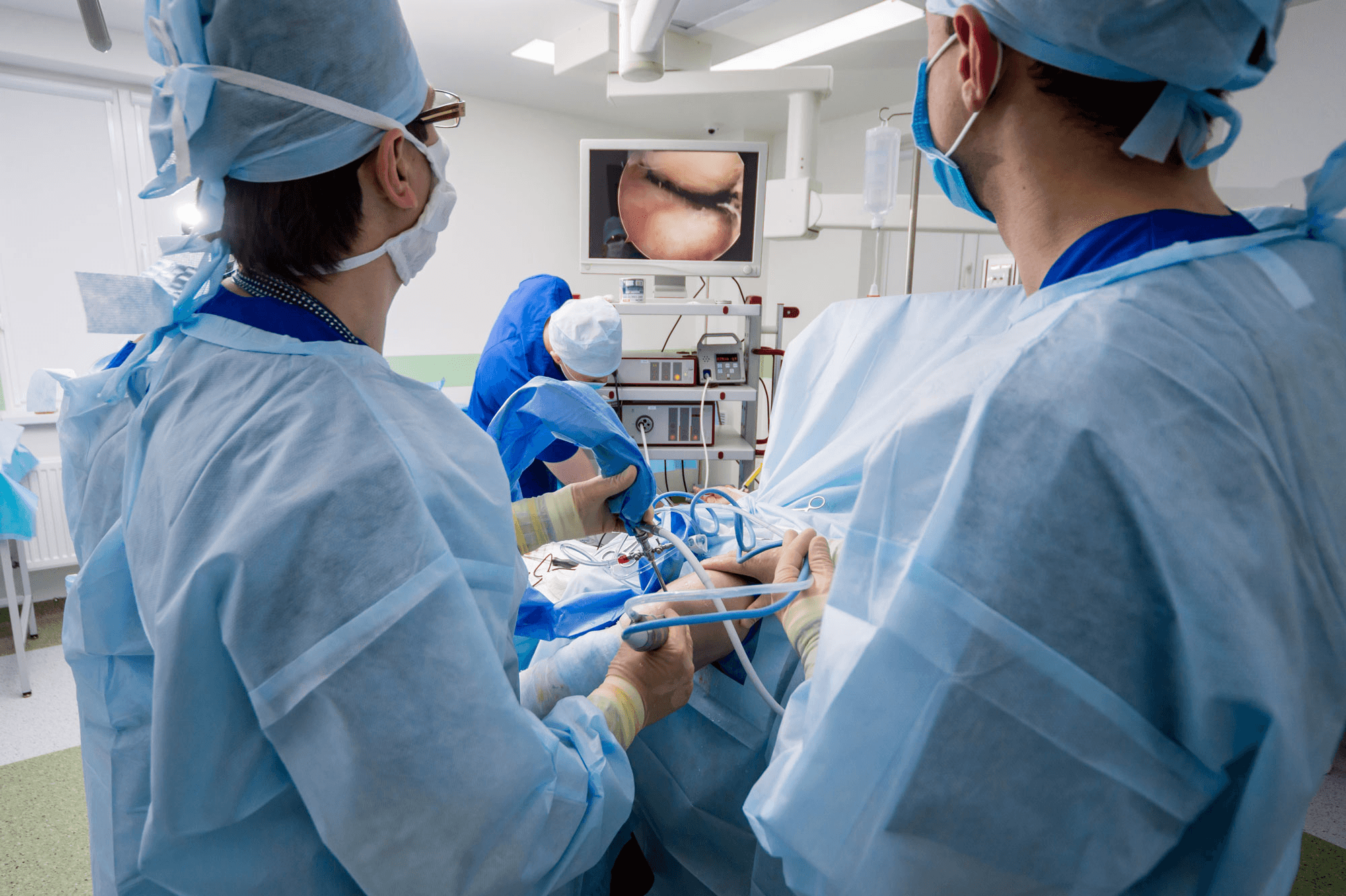
Here is the point most guides miss. The same gel can be given in two ways, and they are not recovered from in the same way. One is an ultrasound-guided injection in an outpatient room under local anaesthetic. The other is keyhole (arthroscopic) implantation, the Liquid Cartilage route, which is a surgical procedure. Their recoveries differ substantially, and the sections below keep them apart. For the injection in detail, see our injection recovery page.
Professor Paul Lee, an expert in modern cartilage repair, notes that this minimally invasive approach provides an alternative to more extensive surgeries such as microfracture or cell transplantation. Recent research into cartilage-repair materials has found that ChondroFiller has properties that can help joints better handle mechanical stress during healing (Weizel et al., 2020).
How to Prepare for Your ChondroFiller Treatment
Preparation can make a big difference in your healing experience. Healthy daily habits, such as quitting smoking, keeping a balanced diet, and maintaining a healthy weight, can help your body recover whichever route you take.
Before your treatment, you'll have scans and assessments to pinpoint the cartilage injury and agree which route suits you. Share your complete medical history and a list of any medications with your care team, so they can ensure you're set up for a smooth, safe procedure . If you are having the injection, plan for someone else to drive you home, as you should not drive on the day. If you are having keyhole surgery, also plan for time away from work and for crutches at home.
Meeting with a specialist like Professor Paul Lee will help you understand your specific needs and what you can expect from ChondroFiller . The team at MSK Doctors is dedicated to answering your questions and preparing you fully.
What Happens Right After Your ChondroFiller Procedure?
After the injection
The injection takes 30 to 45 minutes in an outpatient room, and you walk out the same hour. The local anaesthetic wears off in about six hours. There is no brace, and no crutches or walking aids are needed unless your medical team specifies otherwise. Your dressing comes off the next day and the site is kept dry for 48 hours. Days 1 to 3 are quiet, with ice as needed, and some soreness or swelling around the joint is normal. Please do not drive on the day.
Weeks 1 and 2 are for integration: aim for about half your usual joint activity, and avoid massage or deep heat for the first week. Weeks 3 to 6 are the repair phase, with a graded return to normal activity. A free telephone review at six weeks checks your progress.
After keyhole surgery
Some soreness or swelling around the treated joint is also normal here. To protect your cartilage as it heals, the joint is protected in a brace or splint for the first 48 hours. Soon after, you'll start gentle movement exercises, often with the support of a specialised device, to promote healing without stressing the new cartilage . Follow your surgeon's instructions on pain relief and infection prevention carefully.
For about six weeks you will be partially weight-bearing. This means using crutches or other supports, as directed by your care team. Your specialists will provide a detailed aftercare plan, and regular check-ins with the MSK Doctors team will keep your recovery on track.
Long-Term Recovery: Getting Back to Daily Life and Beyond
Injection route
After week six, the aim is to keep building activity at a sensible pace. Cartilage improvement is expected over 6 to 12 months, and a 12-month MRI with Professor Lee shows how the repair is maturing. Physiotherapy is optional: some people use it to structure a return to sport, but it is not required.
Keyhole route
Patience is an important part of recovery here. After the partial weight-bearing phase, weight is rebuilt over about six to eight weeks, and cycling and swimming resume steadily as strength returns. Physiotherapy forms part of this route, helping you rebuild strength and flexibility with careful, progressive exercises to support the healing cartilage. High-load or contact sport, such as running or jumping, is held back until around six months, and most sports are back at around 12 months, giving your new cartilage time to mature and become resilient. This is the manufacturer's rehabilitation, also followed in arthroscopic series such as the hip technique described by Perez-Carro et al. (2021).
On either route, regular follow-up with your specialist helps monitor your progress, adjust your plan as necessary, and keep your joint healthy for the long term.
Conclusion and Disclaimer
Taking the right steps before and after your ChondroFiller treatment can significantly improve your results, and knowing which route you are on is the first step. With the guidance of Professor Paul Lee and the team at MSK Doctors , you'll be supported throughout. For specific advice about your situation, always speak with a qualified healthcare professional.
References
Breil-Wirth, A., von Engelhardt, L., Lobner, S., & Jerosch, J. (2016). Retrospective study of cell-free collagen matrix for cartilage repair. , , . https://doi.org/10.3238/oup.2016.0515-0520
Perez-Carro, L., Rosi Mendoza Alejo, P., Gutierrez Castanedo, G., Menendez Solana, G., Fernandez Divar, J. A., Galindo Rubin, P., & Alfonso Fernandez, A. (2021). Hip Chondral Defects: Arthroscopic Treatment With the Needle and Curette Technique and ChondroFiller. , , . https://doi.org/10.1016/j.eats.2021.03.011
Weizel, A., Distler, T., Schneidereit, D., & Friedrich, O. (2020). Complex mechanical behavior of human articular cartilage and hydrogels for cartilage repair. Acta Biomaterialia, , . https://doi.org/10.1016/j.actbio.2020.10.025
Frequently Asked Questions
- ChondroFiller uses a collagen scaffold to encourage your body's own stem cells to regenerate cartilage. Professor Paul Lee at MSK Doctors offers it as an ultrasound-guided injection or, for larger or more complex defects, through keyhole implantation. Both use the same gel, but their recoveries differ substantially.
- Preparation includes healthy habits, sharing your medical history, and undergoing scans. Your team, led by Professor Paul Lee, agrees which route suits you. If you are having the injection, arrange for someone else to drive you home, as you should not drive on the day. For keyhole surgery, plan for crutches and time away from work.
- After the injection you walk out the same hour, with no brace and no crutches unless your medical team specifies otherwise; the dressing comes off next day and the first three days are quiet. After keyhole surgery the joint is protected in a brace or splint for the first 48 hours, followed by about six weeks of partial weight-bearing with crutches or supports.
- After the injection: about half your usual joint activity in weeks one and two, a graded return in weeks three to six, a free six-week telephone review and a 12-month MRI; physiotherapy is optional. After keyhole surgery: weight rebuilt over about six to eight weeks with physiotherapy, steady return to cycling and swimming, high-load sport from around six months and most sports at around 12 months.
- Professor Paul Lee is renowned for his expertise in cartilage repair, and MSK Doctors delivers patient-centred care on both the injection and keyhole routes. Their experience with ChondroFiller means you receive knowledgeable advice on which route suits you and tailored support throughout your recovery.
Next steps
Where to go from here
These routes are selected from the topic and purpose of this article. They are guidance, not a diagnosis or treatment recommendation.
Recovery route
Explore recovery guidance
Continue with practical recovery and patient-journey information.
Talk to the team
Book a free discovery call
A non-medical call with the team to understand services and choose the right booking route.
Legal & Medical Disclaimer
This article is published by Liquid Cartilage for general information and education only. It does not constitute medical advice, diagnosis, or treatment.
Always seek personalised advice from a qualified healthcare professional before making decisions about your health. Liquid Cartilage accepts no responsibility for errors, omissions, third-party content, or any loss, damage, or injury arising from reliance on this material.
If you believe this article contains inaccurate or infringing content, please contact us at [email protected].